Showing posts with label Vascular. Show all posts

Sunday, 7 August 2016

Procedure: Femoral artery and vein catheter insertions

These two videos demonstrate the ultrasound techniques for femoral artery and vein catheter insertions.

First, the femoral artery catheter insertion:


Second, the femoral vein catheter insertion:


These are both very similar, since you will be locating both the femoral artery and vein when you perform either procedure. 

Localization of both of these structures is key when performing these procedures. It is important to remember the anatomy of the femoral region. The femoral artery runs laterally to the femoral vein and medially to the femoral nerve as in the figure below.
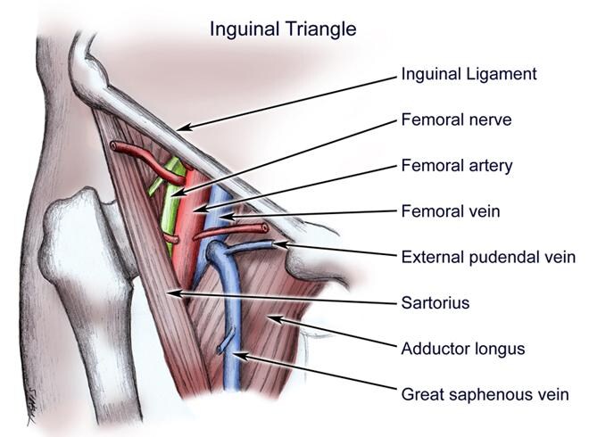


However, anatomical variants do exist, so this is not always the case. To further help with localization of your chosen vessel, remember that that veins are compressible on ultrasound, while arteries are not. 


Sunday, 31 July 2016

Procedure: Radial Artery Catheter Insertion

Although radial artery catheter insertion can be done as a palpation-guided procedure, it can sometimes be difficult to perform successfully. This video provides a general overview on how to perform this procedure with ultrasound guidance.


When performing this procedure, the radial artery appears as an anechoic circle that is pulsatile. Unlike the surrounding veins, the radial artery should not be compressible. If you are having difficulty finding the radial artery, the use of colour doppler can help with localization.


Friday, 27 May 2016

Procedure: Internal Jugular Central Line Insertion

One of the procedures where ultrasound is useful is for central line insertion into the jugular vein. In fact, ultrasound guidance for internal jugular line placement is considered standard of care.


The ultrasound portion of the technique begins at 3:43. You can use the transverse (at 5:00) or longitudinal view (at 5:05) to visualize the tip of the needle as it is inserted into the jugular vein.

When inserting the needle, you should assure the following:
- The internal jugular is centered on the ultrasound image
- The ultrasound probe is perpendicular to the needle's insertion angle is using the transverse view
- The needle's insertion point is at the middle of the ultrasound probe

As mentioned in the video, you should also confirm that the wire is correctly positioned in the jugular vein before proceeding with dilating.